
Skull base meningioma: why nerve function and anatomy matter in a Shanghai review
skull base meningioma function assessment Shanghai
When a skull-base meningioma sits near vision or hearing nerves, blood vessels or the brainstem, its size alone cannot settle treatment. A Shanghai skull-base review can combine symptoms, serial scans and neurological function to compare observation, surgery and radiation with the abilities that matter most to you in mind.
By eastmedgo editorial ·
On this page
Separate a tumor type from its anatomical address
Meningioma describes a tumor arising from the membranes around the brain and spinal cord. Skull base describes a location that can contain several different tumor types. The two terms are not interchangeable. MRI may strongly suggest a meningioma, while tissue examination, when obtained, establishes the pathological diagnosis and grade. Ask whether the report describes a presumed imaging diagnosis or a confirmed one, and keep any previous tissue results available. An apparently benign tumor can still cause important problems through its position; a reassuring label does not replace assessment of pressure on surrounding structures.
Ask what the tumor touches, displaces or surrounds
The skull base contains pathways for vision, hearing, facial movement, swallowing and other functions, alongside major vessels. Tumors close to the optic nerve raise different concerns from those near the brainstem or vessels at the petroclival region. Review original MRI images rather than relying only on a largest-diameter measurement. Ask the specialist to identify the structures at risk, any pressure effect and the relationship that most limits treatment. A second opinion should describe these findings in understandable language. “Difficult location” is less useful than a precise explanation of which function or vessel makes a proposed intervention hazardous.
Sources and references: [1] [2] [3]
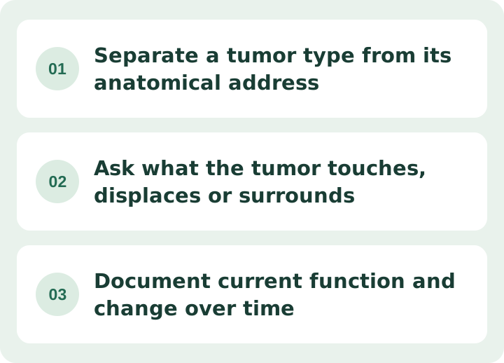
Document current function and change over time
The clinical picture includes vision, hearing, balance, strength and other neurological findings. Depending on the location, formal eye or hearing tests may provide a baseline that a routine scan cannot. Compare scans over time to distinguish a single size measurement from demonstrated growth, and record when symptoms began or changed. Existing impairment and the risk of further loss affect the balance between watching and intervening. Ask which symptoms are plausibly related to the lesion and whether a functional change increases urgency even if growth looks modest. Sudden weakness, a new seizure or rapid major visual change needs urgent local assessment.
Monitoring is an option for selected stable situations
Some small, slow-growing meningiomas without symptoms can be followed with imaging and clinical review. This does not mean every small skull base lesion can safely wait. Proximity to sensitive structures, documented growth, symptoms and overall health influence the recommendation. Ask for the imaging schedule, the functional tests to repeat and the findings that would trigger treatment discussion. The plan should identify who will compare scans and how you will report new symptoms between visits. If tissue has never been obtained, the report should preserve the distinction between a presumed meningioma and a histologically established grade.
Define the goal as safe tumor control and function preservation
Surgery may be considered for a growing or symptomatic tumor, but complete removal is not always the safest goal when critical nerves or vessels are closely involved. A planned residual portion may lead to surveillance or a radiation discussion, depending on pathology and other findings. Ask what the surgeon aims to remove, what may deliberately remain and why. Discuss bleeding, infection and location-specific neurological risks, including possible visual or other functional loss. An endoscopic or smaller-opening approach is suitable only for selected anatomy; the name of the approach does not guarantee a lower risk in your case.
Understand what radiation can and cannot replace
Focused radiation may be considered when surgery is unsuitable or for residual tumor in selected circumstances. A single-session radiosurgery plan and a fractionated course are different options, with suitability affected by tumor size and proximity to sensitive structures such as the optic pathway. Radiation aims to control tumor growth; it is not equivalent to physically removing a mass or obtaining tissue for diagnosis. Ask how the team balances control with the risk of injury to nearby structures, how response will be assessed and whether continued observation is a reasonable alternative. Follow-up remains necessary after treatment.
How Shanghai skull-base teams compare observation, surgery and radiation
Huashan Hospital, Fudan University brings together skull-base surgery, microsurgical and endoscopic approaches and multidisciplinary neuro-oncology, described in its hospital specialty overview. For a patient with meningioma, Shanghai’s advantage is a shared question for surgery, radiation and functional testing: what might be lost by waiting, what nerves or vessels shape an operation, and which abilities might treatment preserve or affect? Read about Meningioma and skull base tumors, then bring serial MRI and vision or hearing assessments to compare the purpose and cost of each option.
Sources and references: [4]
Plan the consultation around the function you want to protect
Tell the team how changes affect reading, work, balance, communication and daily independence. Ask for separate estimates for review, imaging, functional testing, surgery or radiation, admission and rehabilitation. Confirm which follow-up scans and specialist tests can occur at home, who will review them and how unexpected deterioration will be handled.
eastmedgo knows the Chinese medical system and can organise serial MRI and changes in vision, hearing and neurological function, connect you with skull-base surgery and radiation specialists, and coordinate the observation-versus-treatment decision with follow-up at home. See eastmedgo services, then contact eastmedgo with serial MRIs and changes in vision or hearing to ask when to observe and when surgery or radiation merits review.
Common questions
Does a small tumor near the optic nerve always need surgery?
No. Its relationship to the nerve, visual function, growth and the risks of each option must be assessed together. Small size alone neither mandates surgery nor establishes that observation is safe.
Is leaving part of a meningioma always a failed operation?
No. Deliberately limiting removal may protect a vital structure. The significance of residual tumor depends on location, grade and the follow-up plan, which may include observation or radiation. Ask what was planned and what the postoperative findings show.
Can MRI tell me the exact tumor grade?
Imaging can suggest a meningioma and describe its behavior, but a pathological grade requires tissue assessment. Not every patient needs a biopsy immediately; the team weighs the value of confirmation against the risks and the management plan.
Sources and references: [1]